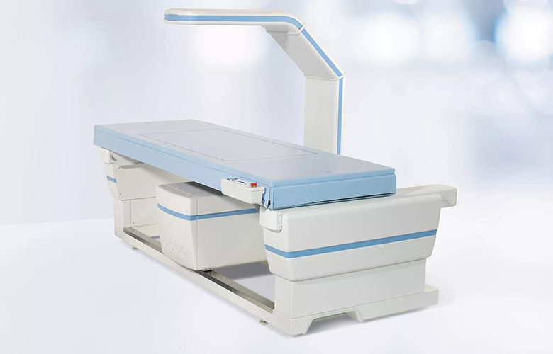
Bone densitometry, also known as dual-energy X-ray absorptiometry (DXA), is a specialised imaging technique used to measure the density of bones in the body. This non-invasive procedure is commonly performed to assess bone mineral density and diagnose conditions such as osteoporosis.
By utilizing low-dose X-rays, a bone densitometry scan can accurately evaluate the strength and quality of bones, helping healthcare providers identify the risk of fractures and tailor appropriate treatment plans for their patients.
During a bone densitometry scan, the patient lies on a table while a scanner passes over the body, typically focusing on the hips, spine, or forearm. The scan generates detailed images and provides precise measurements of bone mineral content, offering valuable insight into the overall bone health of the individual.
This information is crucial in early detection of bone loss and guiding interventions to prevent fractures and promote bone strength. Bone densitometry scans are quick, painless, and essential in managing bone health, making them a vital tool in assessing and monitoring skeletal conditions in patients of all ages.


