Specialised Breast & Ultrasound Center

Breast Center

3D Mammography

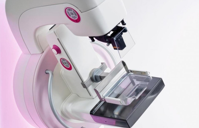
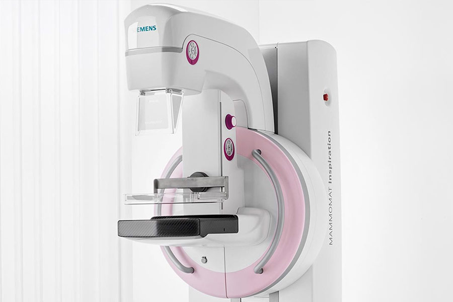
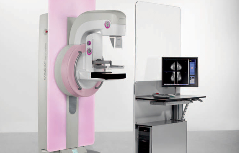
At Diagnosis Breast Centre we offer the latest, state of the art equipment from Siemens Healthineers, the industry leader in advanced breast imaging offering 50 degrees wide-angle Tomosynthesis which is the widest angle in the market for enhanced diagnostic capabilities and outstanding image quality.


All  mammograms performed at our center include 3D Tomosynthesis. Mammograms performed using 3D Tomosynthesis increase diagnostic efficacy of the study and reduce false positives. This means a more accurate examination with less room for error.

Multiple studies show that 3D mammography improves cancer detection. 3D mammograms can detect up to 40% more cancers than 2D mammograms, simply we are able to find cancers that we would not have been able to find with 2D.


Mammograms can be performed as a Screening Mammogram, or a Diagnostic Mammogram. Screening Mammograms are performed on patients who are asymptomatic, and who fall within specific age guidelines. Diagnostic Mammograms are generally performed on symptomatic patients, or patients who have a positive finding on a screening mammogram.

Diagnostic Mammogram

Diagnostic Mammograms are ordered for patients who have had an abnormal screening mammogram, or who have signs or symptoms of breast cancer. Unlike screening mammograms, diagnostic mammograms are not performed on an annual basis. They are typically localized to the breast of interest and include additional views not traditionally performed during a screening mammogram. Reasons to have a diagnostic mammogram may include:

Feeling a lump in the breast or axilla
Breast pain
Nipple discharge
Breasts that have changed size or shape
Breast imaging abnormality

Breast Ultrasound

Breast ultrasound is an imaging test that uses sound waves to look at the inside of your breasts.

Breast ultrasound is not usually done to screen for breast cancer. This is because it may miss some early signs of cancer. An example of early signs that may not show up on ultrasound are tiny calcium deposits called microcalcifications.

Breast Elastography

Breast ultrasound elastography provides information on breast lesions in addition to conventional ultrasonography (US) and mammography. Elastography provides a noninvasive evaluation of the stiffness of a lesion. In our breast center we can provide both strain and shear wave elastography.

Breast Biopsies and Interventions (core biopsies).


Image-guided percutaneous breast biopsy (Core & FNA) is a simple, safe, accurate, and almost painless method of establishing a tissue diagnosis of breast abnormalities.

In breast biopsy we use special needles with which we remove a small sample of tissue and send it for analysis to a specialised laboratory.

Prior to surgery for removal of an abnormality in your breast that cannot be easily felt, the radiologist will place a hookwire to guide the surgeon during operation.

A hookwire gives the surgeon an accurate location of the lump prior to surgery.

A clip placement is a marker placed in the breast, lymph node, or both. It locates and marks an area of concern that a doctor found on a mammogram or ultrasound. After chemotherapy shrinks the tumour and lymph node, the doctor can still find these areas of concern and remove them.

Breast biopsies (core biopsies), hookwire localisation and clip marker placement are carried out using local anaesthetic to numb the breast at the area of interest.