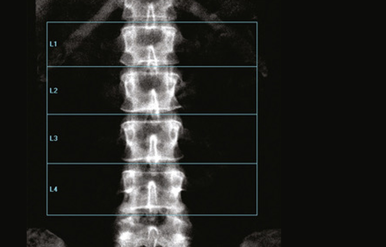
A dual-energy X-ray absorptiometry (DEXA) scan measures the density and mineral content in bone, most often in the hip or lower spine. It is the most accurate method of determining bone density and potential problems related to bone loss.
Osteoporosis resulting in bone fractures is responsible for thousands of injuries in the elderly that require long-term care confinement. Osteoporosis, or bone loss, is commonly associated with post-menopausal women. However, it can also affect men. Therefore, it is important for everyone with risk factors—such as being post-menopausal without estrogen supplementation, patients who are smoking or have smoked, and those individuals taking certain medications such as Prednisone or other steroids, and other factors—to have a DEXA scan.
DEXA scan is a quick and painless procedure. You simply lie on a flat padded table, the arm of the machine passes over your body, and the scan takes approximately 10-15 minutes. You remain clothed during the scan. You will be able to communicate with the technologist during the procedure. Results from the test will be sent to your doctor.
A DEXA scan is painless and non-invasive, with very little exposure to radiation—less than one-tenth of what is used in a chest X-ray.
At our facilities, we have DEXA scanning equipment with the latest software for the most accurate diagnosis. This enhanced form of X-ray is more accurate than a plain X-ray test and provides the only way to accurately diagnose and monitor a patient with significant bone loss. This test is a valuable tool for diagnosing osteoporosis, which often has no symptoms until you suffer a fracture. A Bone density scan can diagnose the disease at its earliest stages, which means you can begin receiving treatment to protect your bones sooner.

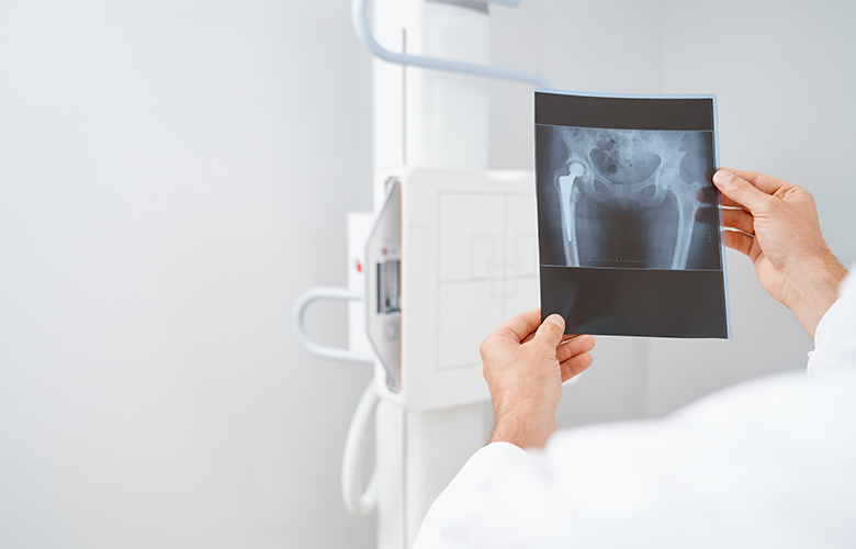
X-ray is the oldest and most frequently used form of medical imaging. It is also the fastest, and easiest and most economical way for a physician to view and assess broken bones.
It can also be used to diagnose and monitor the progression of diseases, including heart disease, and cancer. Unlike other forms of radiation, X-rays can easily pass through body tissue, making it possible to provide images of internal structures without performing surgery.
During the procedure, electromagnetic radiation passes through the body onto “film” (now digitized and displayed on a computer screen). Dense structures such as bone absorb most of the radiation and appear white on the digital image. Structures that are less dense like air appear black. Everything in between appears a different shade of gray.
Digital X-rays are used to diagnose a wide range of illnesses and injuries, including musculoskeletal injuries, cancer, lung infection, abdominal pain, sinus disease, spinal problems and other abnormalities.
You may be asked to stand or lie down on an examination table, depending on the part of the body to be examined. You will be able to communicate with the technologist during the procedure.
There is little reason to worry about the small amount of radiation you will be exposed to when you receive a digital X-ray.
We use the most advanced technology to deliver premium healthcare. Digital imaging gives us many advantages in handling your exam, not the least of which is faster communication of results to your doctor. Digital imaging also allows your medical team to collaborate, if necessary, and to immediately compare previous exams with current ones, so that your health is properly monitored.
